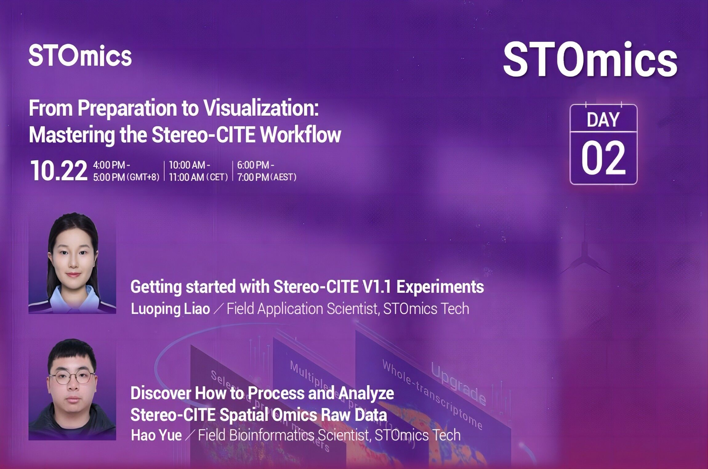
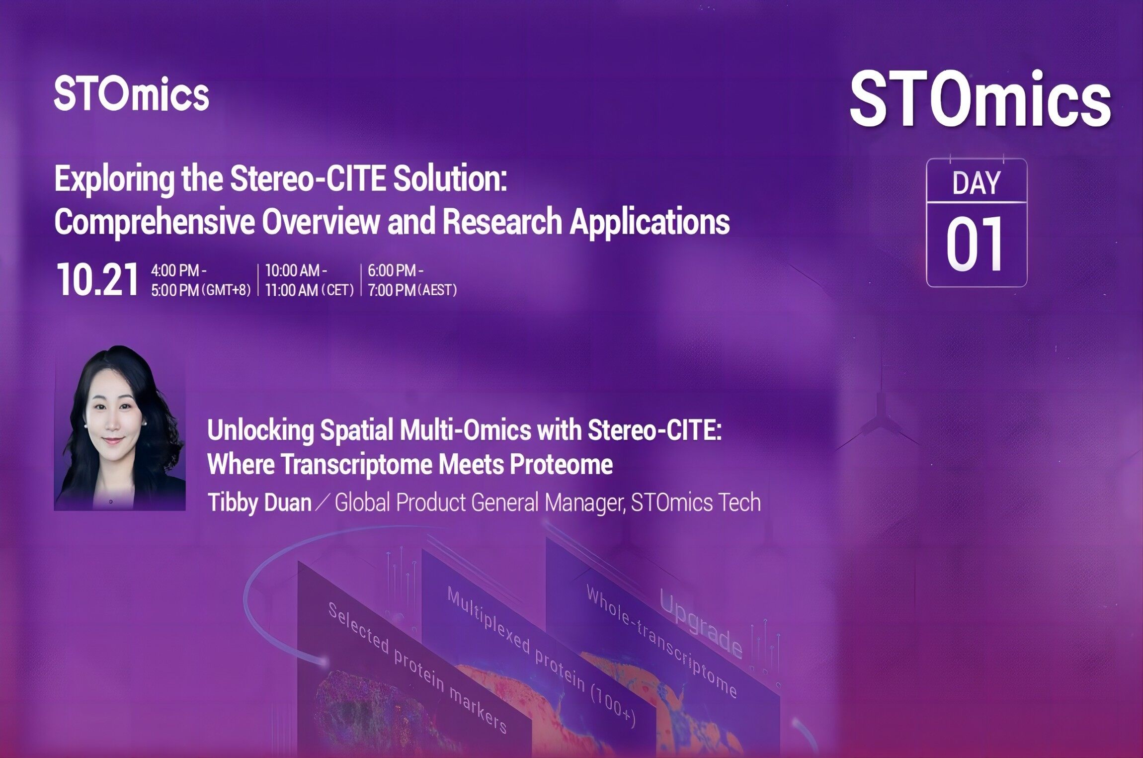
Lab story that mattered — the moment I stopped assuming everything was fine
I remember the day clearly: in July 2023 I ran a 10 mm×10 mm mouse hippocampus section on stereo-seq and the downstream mapping looked off, so I checked the spatial transcriptomics sample results right away. The stereo-seq sample gallery showed examples that matched (mostly) what I was seeing — uneven mRNA capture and weird gaps in gene expression maps, lah. Scenario: bench run with freshly sectioned tissue; data: 27% spot dropouts on one slide; question: how much of our biological signal is just method noise that misleads interpretation? I say this because I’ve been doing spatial work for over 18 years and small biases accumulate — very quickly they change conclusions.
I’ll be frank: classic fixes like cranking up sequencing depth or rerunning tissue sections are blunt. I tried higher read depth in a June run at a central lab in Singapore and found diminishing returns — more reads, but the same spatial artefacts. That taught me that the problem often sits before sequencing: poor tissue adhesion, suboptimal barcode arrays, or uneven permeabilisation can wreck a whole dataset. I use the stereo-seq sample gallery as a quick sanity check now; it’s not gospel, but it helps me spot familiar artifact patterns fast (very practical). Next, I mapped which steps tend to fail and why — the reasons surprised me.
What were the hidden pain points?
Forward-looking fixes — practical checks and measurable selection criteria
I shifted to a forward-looking approach: identify where the workflow leaks signal, then choose tools that give measurable control. First, I validate tissue handling: cold ischemia time recorded (I log timestamps — e.g., 11:20 AM cut, 11:35 AM frozen) and I noticed runs with >15-minute delay showed 10–30% lower mRNA capture. Second, I audit the barcode arrays and surface uniformity; uneven arrays give you spatial bias that’s impossible to fix later. Third, I benchmarked library prep—certain chemistries tolerate degraded RNA better. I checked these against examples in the spatial transcriptomics sample results to see real-case variance, and that comparative look saved me several wasted runs — true story, saved S$3k in reagents on one failed experiment.
Technical note: I use terms like stereo-seq, barcode arrays, mRNA capture and gene expression mapping in daily troubleshooting. Those are not buzzwords — they point to concrete control points. If you want a quick checklist (I keep one taped to my bench): 1) timestamp tissue handling and aim for <10 min to freezing, 2) run array uniformity QC before tissue placement, 3) pilot low-depth sequencing to check spatial consistency before committing to deep runs. These metrics make supplier claims testable. Short interruption — sometimes you must toss a slide and move on. Then recalibrate. The result: fewer surprises, and more interpretable spatial maps.
What’s Next
To choose a solution wisely, I recommend three evaluation metrics — easy to measure, high impact: 1) spatial fidelity score (compare known marker localization versus mapped signal), 2) effective spot recovery rate (percent of spots passing QC post-sequencing), and 3) batch-to-batch variance in mRNA capture. I stick to these when evaluating kits or a new protocol; they tell me quickly whether a change is meaningful. I’ve used them to compare two surface chemistries in March 2024 — one showed 12% better spot recovery and saved us repeat work. So, measure, don’t guess. I keep testing and sharing findings with my team; we learn faster that way. Final note — small pre-run checks beat massive post-run troubleshooting, trust me. For reference and examples, have a look at the stereo-seq sample gallery and the linked galleries from stomics for real-world comparisons. stomics